Lesson 2 ·Class 10 · Sense Organs · Label Lab
The Eyeball's Three Layers and the Retina
Sclerotic, choroid and retina — with the yellow spot and the blind spot
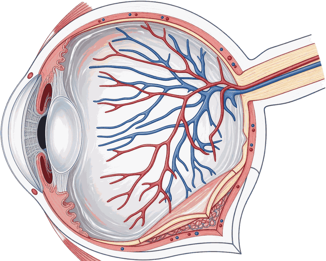
Tap any part of the diagram to learn its name and its job. Try: List which layer does what: protection, nourishment, light detection.
Free play — explore the diagram, or switch to "Label it yourself" and place every label. Switch to when you're ready to test yourself.
Outside in, the wall of the eyeball is sclerotic, then choroid, then retina. The yellow spot is the area of BEST vision; the blind spot beside it, where the optic nerve leaves, has no sensory cells at all.

What this lesson covers
What you do
- List which layer does what: protection, nourishment, light detection.
- Recall that rods hold rhodopsin (visual purple) and cones hold iodopsin (visual violet).
- Explain why you move your eyes from word to word while reading instead of taking in a whole line at once.
The parts you label
- Sclerotic layer (outer) — Tough white fibrous tissue forming the first of the three concentric layers. It maintains the shape of the eyeball and protects it.
- Choroid layer (middle) — Richly supplied with blood vessels to nourish the eye, and packed with the black pigment melanin which stops light rays reflecting and scattering inside.
- Retina (inner) — The innermost light-sensitive layer. It contains rods, which work in dim light and hold rhodopsin, and cones, which need bright light and give colour vision.
- Yellow spot (macula lutea) — The spot at the back of the eye with the maximum number of sensory cells, especially cones. It is the region of brightest and of colour vision.
- Optic nerve — Nerve fibres from all the sensory cells converge and leave the eyeball here, at the blind spot, and carry impulses to the visual area of the cerebrum.
- Vitreous humour — The transparent jelly-like fluid filling the large chamber behind the lens. It keeps the eyeball in shape and protects the retina and its nerve endings.
Challenges to clear
- Tap the layer that both nourishes the eye and blackens it against stray reflections.
- Tap the area of best vision, crowded with cones.
- Place every label correctly.
Hint
Outside in, the wall of the eyeball is sclerotic, then choroid, then retina. The yellow spot is the area of BEST vision; the blind spot beside it, where the optic nerve leaves, has no sensory cells at all.