Lesson 7 ·Class 10 · The Circulatory System · Label Lab
Heart Structure — Chambers, Valves and Major Vessels
Four chambers, four valves, five great vessels.
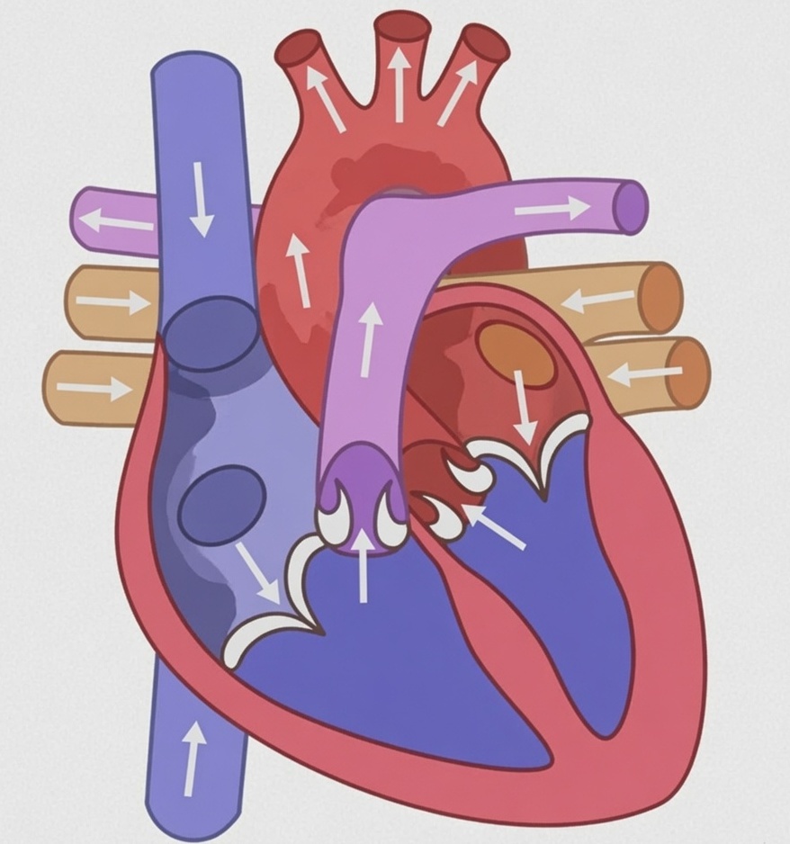
Tap any part of the diagram to learn its name and its job. Try: Cover the labels and say aloud which two vessels bring blood in and which two carry it out.
Free play — explore the diagram, or switch to "Label it yourself" and place every label. Switch to when you're ready to test yourself.
Learn the four valves as two pairs. Tricuspid, three cusps, right atrium to right ventricle; bicuspid or mitral, two cusps, left atrium to left ventricle; and pocket-shaped semilunar valves, three each, at the mouths of the pulmonary artery and the aorta. The chordae tendineae are the parachute cords that stop the cuspid flaps turning inside out.

What this lesson covers
What you do
- Cover the labels and say aloud which two vessels bring blood in and which two carry it out.
- Explain why the heart, handling the whole blood supply, still needs its own coronary arteries to feed its walls.
The parts you label
- Vena cava — Two great veins empty into the right atrium. The anterior or superior vena cava brings deoxygenated blood down from the head, chest and arms; the posterior or inferior vena cava brings it up from the abdomen and legs. Both carry blood towards the heart, which is what makes them veins.
- Right atrium — The upper right chamber, receiving deoxygenated blood from both venae cavae. Its walls are thin because it only has to push blood into the ventricle just below it. The pacemaker or sino-atrial node sits in this wall, near the opening of the superior vena cava.
- Right ventricle — The lower right chamber. Blood reaches it through the right atrio-ventricular valve, which has three flaps and is therefore called the tricuspid valve. Its wall is muscular, but thinner than the left ventricle because it only has to pump as far as the lungs.
- Pulmonary artery — Leaves the right ventricle through three pocket-shaped pulmonary semilunar valves and divides to enter both lungs. It is the one artery in the body carrying deoxygenated blood — the classic exception in every artery-versus-vein question.
- Left atrium — The upper left chamber. It receives four pulmonary veins, two from each lung, bringing oxygenated blood back from the lungs. These are the one set of veins carrying oxygenated blood — the mirror-image exception to the pulmonary artery.
- Left ventricle — The lower left chamber, entered through the left atrio-ventricular valve, which has two flaps and is called the bicuspid or mitral valve. Its wall is the thickest in the heart because it must drive blood to the farthest points of the body, up to the toes and up to the brain against gravity.
- Aorta — The largest artery, arising from the left ventricle past three aortic semilunar valves and carrying oxygenated blood to the whole body. The two coronary arteries branch from its very base to feed the heart muscle itself; a block in one of them causes a heart attack.
Challenges to clear
- Find the chamber with the thickest wall in the heart.
- Now find the only artery that carries deoxygenated blood.
- Label all four chambers and the great vessels.
Hint
Learn the four valves as two pairs. Tricuspid, three cusps, right atrium to right ventricle; bicuspid or mitral, two cusps, left atrium to left ventricle; and pocket-shaped semilunar valves, three each, at the mouths of the pulmonary artery and the aorta. The chordae tendineae are the parachute cords that stop the cuspid flaps turning inside out.